Tales from the Grave: Concha Bullosa
This is a small series that will focus on some of the types of trauma, pathology, interesting variations and degeneration I examined while in Cyprus at the Odyssey Field School. Please note that the images included are NOT from the actual cases that my peers and I worked on. However, the images that are included are accurate representations of the cases analyzed.
Concha Bullosa
In order to better understand what concha bullosa is, I think it may help to breakdown the various components of the feature.
Where on the body does concha bullosa happen? Inside the nose. Your nose is made up of many different thin small bones.
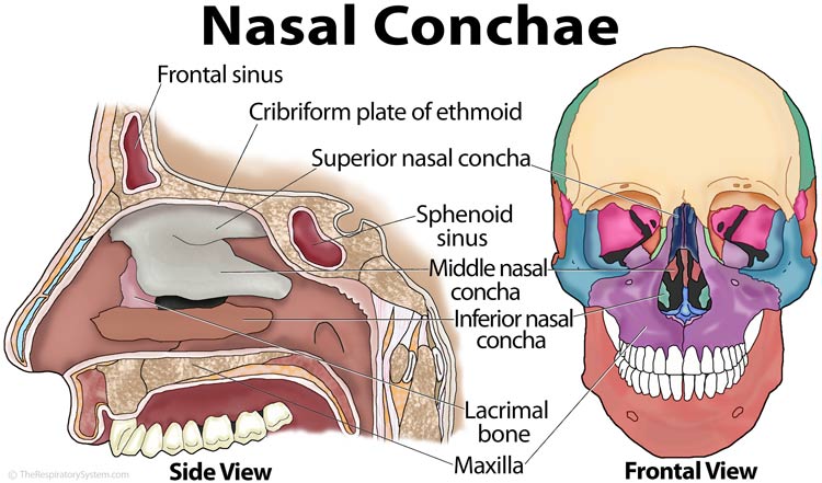
The affected bone is the nasal concha (turbinate) which is a narrow bone that sticks into your breathing passages. “The nasal conchae or turbinates are named this way because they function in a similar way as a turbine, being principally responsible for regulating the air flow during inhalation.”
These turbinates help us with nasal functions like smelling, keeping our respiratory system moist, etc. They can enlarge when irritated. When they do, we might experience issues like sinus pressure and stuffy nose. Irritation can occur from issues like cold, flu, allergies, etc.
Next term!
Pneumatization is a word that means the presence or development of air-filled cavities in the bone. Skeletal pneumaticity is the presence of air spaces within bones. It looks a like a proper bubble made out of bone.
So now, if I provide you with the medical definition of concha bullosa, I hope you can start to make sense of what it is:
“The term ‘concha bullosa’ refers to the pneumatisation of the intranasal turbinates.” Basically, those nasal concha puff out.
Now I want to show you what it looks like in the skull:
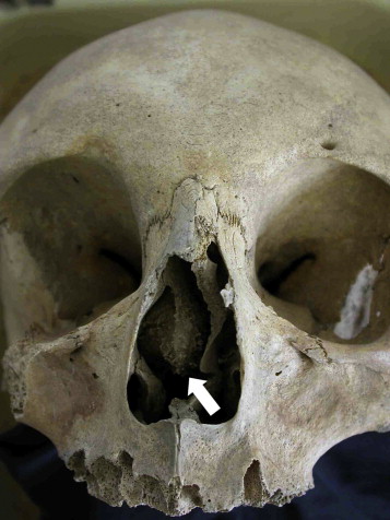
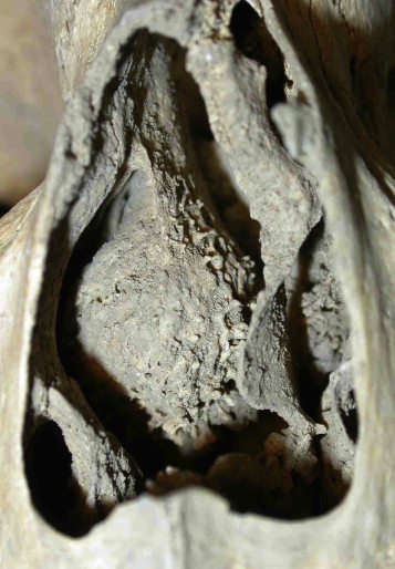
What should be a very flat, narrow, sharp edged bone has the appearance of being puffed up.
We saw this in several individuals during our internship. On one individual, concha bullosa appeared bilaterally, meaning on both sides.
An official CB diagnosis often requires more than analyzing the morphology of the area. This is because it can resemble other, lesser seen conditions, such as fibrous dysplasia. X-rays and CT scans are one way that we can view the internal space of the bone to ensure that we have rightfully ruled out any other conditions.
A person that has concha bullosa may be asymptomatic or may experience a myriad of respiratory issues within their lifetime. It’s also typically observed as an anatomical variation rather than a pathology, but it is a condition still being studied to better understand why it occurs.
When added to a skeletal report and analyzed alongside other data or information about an individual, the presence of concha bullosa may help further or confirm the identity of a person.
Resources:
A case of concha bullosa mucopyocele in a medieval human skull
https://onlinelibrary.wiley.com/doi/10.1002/oa.1137
A case of Concha Bullosa and potentially related evidences. Concha bullosa discovered in the bones of a medieval skeleton from Brentonico, northeast Italy: a case report (PDF)