Seeing Cancer on the Bones
While doing a biological profile on a set of remains, my friend and colleague pointed out some small lesions along the pelvic girdle of our individual. He called over to our professor and in his very poised British accent announced, “we have some lytic lesions here.” Then they both proceeded to help me identify the ways that cancer can appear on the bone.
Up until that point, I had only seen the very pronounced instances of cancer that totally transform and overtake the morphology of a bone. However, the changes that can occur as cancer metastasizes in the bone can range from subtle to pronounced. Bone metastasis happens when cancer cells from their primary location or tumor move to the bone. This movement can cause the bone to change in places on the bone. This destructive change is known as osteolysis. Osteolysis can occur with other diseases, but we’ll focus on cancer in this post. The lytic lesions appear as small holes on the bone. Being able to examine and identify the presence of these types of bone changes can provide valuable health and environmental details about a person and a population when soft- tissue is no longer available.
When the cancer is active these lesions or holes in the occur and affects the stability of the bone. Available medical treatments can lead to the cancer going way, also known as remission. However, the area of the lesion doesn’t fully recover or regenerate normally. The individual may be offered additional treatments or options to help regain some of its lost strength. Yet, the cancer can still leave its imprint, the experience of the disease, remains on the bone. The scaffolding of the bone that is created via osteoblasts doesn’t fully stimulate bone healing in the affected areas.
What does this look like on the bone?
When developing a biological profile from skeletal remains, the recognition and examination of disease is a very important aspect. Shape, size, dimension, manifestation, and how often these lesions can be viewed on the bone helps to determine whether the forensic anthropologist should be considering cancer or some other disease.
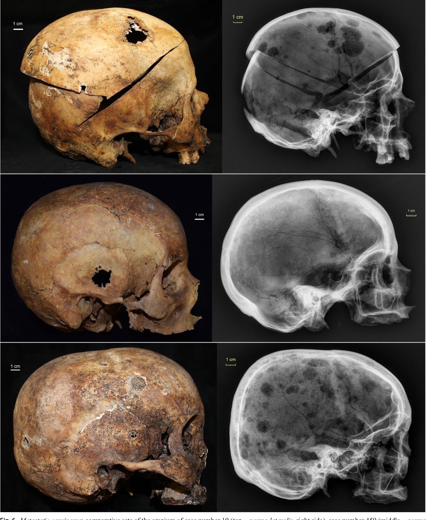
This image shows three crania with evidence metastatic cancer and their corresponding x-rays. These individuals were part of a study examining the presence of cancer on dry bone. The photographs to show some of the small-holed lesions described above. You’ll also see the x-rays show a lot more skeletal changes than what we can see in the picture.
An important thing to keep in mind is diagnosing cancer in human remains can be difficult even when there may be a presence of these types of lesions. X-rays and scans are vital tools for supporting initial detections of suspected cancer.
Cancers whose primary location is the bone is pretty rare and noncancerous bone tumors are more common. Depending on care, treatment, how long an individual lives, or if the cancer moves or metastasizes to the bone, we may not see the disease in skeletal remains, especially with the naked eye alone.
Learn more:
Biehler-Gomez, L., Tritella, S., Martino, F., Campobasso, C.P., Franchi, A., Spairani, R., Sardanelli, F., & Cattaneo, C. (2019). The synergy between radiographic and macroscopic observation of skeletal lesions on dry bone. International Journal of Legal Medicine, 133, 1611-1628.
Biehler-Gomez L, Giordano G, Cattaneo C. The appearance of breast cancer metastases on dry bone: Implications for forensic anthropology. J Forensic Leg Med. 2019;61:5-12. doi:10.1016/j.jflm.2018.10.007